
Advanced Oncology Testing: Tracking Cancer Progression Explained
Have you ever wondered how Specialist Health Practitioners in New Zealand can see the invisible shifts within a patient's body in real time? Modern oncology testing tracking cancer progression has fundamentally changed how we approach long-term health outcomes. These sophisticated tools offer a clearer window into the disease landscape than ever before.

Advanced Oncology Testing Tracking Cancer Progression
By utilising these advanced diagnostic methods, clinicians can now observe subtle changes with unprecedented precision. This shift towards proactive cancer progression monitoring empowers families to navigate complex journeys with greater confidence and clarity. Understanding these breakthroughs is the first step in mastering your own path to wellness.
Key Takeaways
Modern diagnostics allow for highly accurate observation of disease behaviour.
New Zealand Specialist Healthcare providers (e.g. NatuneHeal) are adopting these tools to improve patient outcomes.
Regular assessments help clinicians tailor therapies & treatments to individual needs.
Families gain better insight into the recovery process through data-driven results.
Advanced methodologies reduce uncertainty during the management of long-term conditions.
Understanding the Role of Oncology Testing Tracking Cancer Progression
For patients across New Zealand, the ability to monitor cancer activity in real-time is a game-changer. Modern oncology testing tracking cancer progression allows specialist teams to move beyond reactive measures. By identifying subtle biological shifts, clinicians can adjust therapy and treatment strategies before a condition becomes clinically apparent.
The Importance of Early Detection and Monitoring
Effective cancer diagnosis relies on the integration of advanced screening tools that detect abnormalities at the molecular level. Regular oncology screening provides a baseline that helps Specialist Health Practitioners (e.g. Integrative Oncology Practitioners) distinguish between stable conditions and active disease growth. This proactive approach ensures that patients receive the most appropriate care at the right time.
Consistent tumour monitoring is essential for evaluating how well a specific therapy or treatment plan is working. When we track these markers, we gain a clearer picture of the patient's health journey. This level of insight is fundamental to improving long-term survival rates and overall quality of life.
How Pathology Testing Supports New Zealand Patients
High-quality pathology testing serves as the backbone of modern clinical oncology in New Zealand. Local laboratories utilise state-of-the-art methods to process complex samples, providing the data required for precise tumour monitoring. These results empower both the patient and the specialist to make informed decisions throughout the treatment process.
By leveraging sophisticated laboratory techniques, we can identify changes in a patient's condition with remarkable accuracy. This data-driven framework supports a more personalised approach to cancer diagnosis and ongoing care. Ultimately, robust pathology testing ensures that treatment remains responsive to the evolving needs of every individual.
The Science Behind Circulating Tumour Cells (CTCs)
The journey of neoplasmatic cells through the bloodstream is a critical focus for current oncology research. By studying these cells, clinicians can gain a clearer picture of how cancer behaves within the body. This scientific foundation helps us understand the mechanisms behind disease progression and potential spread.
What are CTCs and Why Do They Matter?
Circulating tumour cells, or CTCs, are cells that have detached from a primary tumour and entered the circulatory system. Monitoring the circulating tumour cells count provides vital information about the aggressiveness of a cancer. These cells act as messengers, offering a real-time look at the tumour's biological status.
Tracking these cells is essential for several reasons:
Early identification of potential metastatic risks.
Evaluating how well a patient is responding to current treatments.
Providing a less invasive alternative to traditional tissue biopsies.
The Process of Malignant Cell Isolation
Capturing these rare cells from a standard blood draw is a remarkable feat of modern science. The process of malignant cell isolation involves separating these specific cells from the millions of healthy blood cells present in a sample. Because these cells are so scarce, highly sensitive technology is required to ensure accuracy.
Once the malignant cell isolation is complete, the concentrated sample is ready for detailed analysis. This allows researchers to study the unique characteristics of the cells that have entered the bloodstream. By maintaining high standards in the laboratory, we ensure that the results provide meaningful insights for clinical decision-making.
Flow Cytometry and Immunophenotyping Explained
After isolation, scientists use advanced tools to identify and classify the cells. Flow cytometry is a powerful technique that allows for the rapid analysis of thousands of cells as they pass through a laser beam. This method is highly effective for determining the specific CTCs count in a patient's sample.
Alongside this, immunophenotyping is used to map the surface markers of these cells. This process helps to distinguish between different types of neoplasmatic cells based on their protein expression. Through the combination of flow cytometry and immunophenotyping, we can build a comprehensive profile of the cancer's behaviour, which is a significant step forward for personalised care in New Zealand.
Utilising Blood Cell Markers for Cancer Identification
Oncology biomarkers analysis has transformed how we detect and monitor various malignancies in New Zealand. By providing a non-invasive way to track disease progression, these blood markers serve as indispensable tools for clinicians. They offer a clear snapshot of systemic health, allowing for more informed decisions during oncology screening.
Common Blood Markers: CEA, PSA, and CA19.9
Standardised testing often relies on specific proteins that indicate the presence of tumours. For instance, carcinoembryonic antigen (CEA) is a vital tool in colorectal cancer screening. Similarly, prostate specific antigen (PSA) and acid prostatic phosphatase are essential for assessing prostate health.
Other markers provide further diagnostic clarity for various conditions. These include:
CA19.9, CA50, and CA195 for gastrointestinal monitoring.
TAG72, a glycoprotein often elevated in epithelial cancers (e.g. ovarian, breast, colon, and pancreatic adenocarcinomas).
Specialised Markers: Thyroglobulin, Calcitonin, and Ferritin
Beyond standard panels, specialised markers offer deeper insights into specific organ systems. Thyroglobulin and calcitonin are critical for the early detection and management of thyroid cancer. These tests help Specialist Health Practitioners track how well a patient is responding to ongoing therapy and/or treatment.
Additionally, ferritin levels are frequently monitored as a systemic indicator. While often associated with iron levels, it can also act as a marker for various inflammatory processes and malignancies. Maintaining a balance in these levels is crucial for overall patient wellbeing.
The Significance of CD Markers in Hematologic Malignancies
Cluster of Differentiation (CD) markers are essential for identifying cells of a hematologic origin. These markers allow specialists to classify cells based on their surface proteins, which is vital for diagnosing a hematologic malignancy marker. For example, CD34 and CD133 are often used to identify stem cells, while CD44 and CD45 help characterise immune cell populations.
In the context of myeloid cell origin disorders, specific markers provide diagnostic precision:
CD20, CD52, and CD33 are key targets for targeted therapies.
CD227 and CD63 assist in tracking tumour-derived vesicles.
By utilising these advanced tools, Specialist Health Practitioners can better understand the unique profile of a patient's condition. This precision ensures that personalised care remains at the heart of the oncology journey.
Advanced Testing: Oncology counts
New diagnostic technologies are changing the way we approach metastatic cancer tracking for patients in New Zealand. By utilising advanced testing platforms, clinicians can now access high-resolution data that reveals how cancer behaves at a cellular level. This level of tumour monitoring is essential for creating personalised treatment plans that adapt to the patient's specific needs.
Oncology counts: Quantifying Circulating Tumour Cells (CTCs)
This test is a powerful tool designed to provide an accurate circulating tumour cells count. By identifying these cells in the bloodstream, Specialist Health Practitioners can better understand the current burden of the disease. Maintaining a precise CTC count allows the clinical team to observe how the body responds to ongoing therapies and treatments.
Oncology tracing: Monitoring Metastatic Potential
While oncology counts focus on the number of cells, oncology tracing provides a deeper look into the broader metastatic potential of the disease. This test helps identify the aggressive nature of the cancer by looking for specific markers that indicate a higher risk of spread. It is a vital component for patients who require proactive tumour monitoring to stay ahead of disease progression.
Distinguishing Between CTCs and Circulating Stem Cells (CSCs)
A critical aspect of modern diagnostics is the ability to differentiate between standard circulating tumour cells and more resilient circulating stem cells. Identifying the CSC count is important because these cells are often responsible for therapy or treatment resistance and recurrence. We use a variety of specific markers to ensure accurate identification:
CD44 and CD133: common markers used to identify the presence of stem-like properties.
CD34 and CD45: essential for identifying hematological stem cell and blast cell marker profiles.
CD227 and CD63: specific indicators that help refine the tumour stem cell marker analysis.
By distinguishing these cell types, clinicians can better tailor their strategies. Understanding whether a patient has a high circulating stem cell count allows for more targeted interventions. This precision is what makes advanced testing so valuable in the fight against cancer.
Onconomics and Oncotrail: Personalised Oncology Insights
Modern oncology is shifting towards highly individualised strategies that focus on the specific behaviour of malignant cells. By leveraging advanced laboratory techniques, patients in New Zealand can now access deeper insights into their cancer progression. This proactive approach allows for tumour growth monitoring that is tailored to the unique biological profile of each individual.
Analysing Tumour Growth and Invasiveness
The use of onconomics and oncotrail represents a significant leap forward in diagnostic precision. These tests rely on malignant cell culture to observe how cancer cells behave in a controlled environment. By measuring the invasiveness of these cells, clinicians can better predict how a tumour might spread and develop over time.
"The future of medicine lies in the ability to tailor therapies and treatments to the individual, ensuring that everyone affected receives the most effective care possible without budget constraints." - Dr David Weller (Certified Integrative Oncology Specialist Practitioner & Molecular Biologist).
The Role of Biological Modifier Substances
A key component of these advanced tests is the evaluation of how a biological modifier substance affects cancer cell viability. These substances can include natural compounds or specific drugs that may inhibit or promote tumour activity. Understanding these interactions helps specialist teams to identify which interventions are most likely to yield positive results for the patient.
Evaluating Chemosensitivity and Targeted Therapies
Personalised oncology goes beyond standard protocols by testing how a patient's cells respond to various therapies and treatments. This may include, for example, assessing the efficacy of tyrosine kinase inhibition, which is a common strategy for blocking the signals that tell cancer cells to grow. Targeted therapies are then selected based on these laboratory findings, ensuring that the therapy and treatment plan is strategically aligned with the cancer's specific biological makeup.
By focusing on these precise metrics, patients and their oncology teams can make informed decisions about their care. This data-driven approach reduces the reliance on trial-and-error methods, providing a clearer path forward during the therapy and treatment journey.
Tracking Specific Cancer Types Through Biomarkers
Modern oncology biomarkers analysis offers a clearer window into how different cancers behave over time. By measuring specific proteins and antigens in the blood, clinicians can gain valuable insights into disease progression. This approach allows for more personalised care plans tailored to the unique molecular profile of a patient's condition.

Tracking Specific Cancer Types Through Biomarker Analysis
Breast Cancer Antigens and Mucin 1
Monitoring breast cancer often involves tracking a variety of specific proteins that indicate tumour activity. Mucin 1 is a key breast cancer antigen that helps clinical teams observe how a patient responds to ongoing therapies. Other markers are also frequently utilised to provide a comprehensive view of the patient's health status.
These markers are essential for detecting subtle changes in the body. Common indicators include:
CA15.3 and CA549 for monitoring tumour burden.
c-erb-b2, which provides data on specific genetic expressions.
TAG-72 and CA72.4, often used alongside markers for colon cancer to ensure accurate tracking.
Prostate Cancer and PSMA Expression
For patients managing prostate cancer, the identification of specific proteins is a game-changer in clinical monitoring. The prostate specific cancer stem cell membrane antigen, commonly known as PSMA, serves as a highly reliable indicator for clinicians.
High levels of PSMA expression often correlate with the presence of active disease. By focusing on this specific marker, specialists can make more informed decisions regarding treatment adjustments. This precision helps in maintaining a proactive stance against the progression of the malignancy.
Lung Cancer Markers: CYFRA 21.1 and SNE
When addressing lung cancer, scientific professionals rely on specialised markers to distinguish between different types of growth. CYFRA 21.1 is a widely recognised marker used to assess the status of epithelial cells. Furthermore, specific neural enolase, or SNE, is critical for identifying a lung neuroendocrine malignancy.
"The use of targeted biomarkers allows us to see beyond the surface of a diagnosis, providing a roadmap for effective, long-term patient therapy and treatment management." - Dr David Weller (Certified Integrative Oncology Specialist Practitioner & Molecular Biologist).
These markers provide the necessary data to track how a tumour responds to therapy and treatment over time. By keeping a close watch on these levels, your healthcare team can adapt your care plan quickly. This ensures that you receive the most effective support throughout your journey in New Zealand.
The Impact of Genetic Mutations on Cancer Progression
At the heart of every cancer diagnosis lies a complex web of genetic mutations that dictate disease behaviour. By identifying these specific changes, healthcare professionals can improve tumour growth monitoring and provide more precise care for patients. These mutations, influenced by the environment to generate a specific phenotype (observable traits or characteristics in an individual), act as the primary drivers of malignancy, influencing how cells divide and survive.
VHL Mutations and Renal Carcinoma
The VHL mut (Von Hippel-Lindau) gene (an alteration of the tumour supressor gene) is a critical factor in the development of certain cancers. When this gene is altered, it often serves as a key renal carcinoma marker for clinicians. Monitoring these changes is just as vital as tracking markers for bladder cancer or liver cancer to ensure early intervention.
BCR-ABL and Acute Leukemia Kinase
In the realm of blood-related malignancies, the BCR-ABL fusion gene is a well-known driver. This mutation is frequently associated with acute leukemia kinase activity, making it a significant leukemia marker for diagnosis. Clinicians often look for specific indicators like CD19, CD99, and the ALK protein to refine their approach.
These markers are essential when evaluating patients with lymphoma or myeloma. By testing for a specific lymphoma related antigen or elevated beta-2-microglobulin, specialists can better understand the disease profile. This level of detail helps in identifying resistant phenotype markers that might otherwise complicate therapy and treatment success.
Understanding Tumour Suppressor Genes and PTEN
A tumour supressor gene acts as a natural brake on cell division, preventing uncontrolled growth. One of the most important examples is PTEN (phosphatase and tensin homolog), which plays a major role in regulating cellular health. When this gene is lost or mutated, the body loses its ability to trigger oncogen apoptosis, or programmed cell death, in damaged cells.
Understanding these genetic pathways allows for a more personalised approach to oncology in New Zealand. By focusing on these molecular triggers, your healthcare team can better predict how a tumour might respond to therapy and treatment. This proactive strategy remains a cornerstone of modern, effective cancer management.
Immunomodulation and the Tumour Microenvironment
The tumour microenvironment acts as a complex ecosystem where immunomodulation dictates how cancer progresses. This environment is not merely a collection of cells but a dynamic space where various signals determine survival and growth. By studying these interactions, clinicians can better understand the mechanisms that allow malignant cells to evade the body's natural defences.
Cytokines, VEGF, and Angiogenesis
Cancer cells often manipulate their surroundings by secreting cytokines to promote their own survival. These signalling proteins, alongside growth factor receptors like EGF-R and IGF-R, create a supportive niche for the tumour. Furthermore, the release of VEGF, FGF, and PDGF triggers angiogenesis, the process of forming new blood vessels to supply the tumour with nutrients.
This vascular network is essential for the tumour to expand beyond its original site. By monitoring these markers, including the endothelial cell membrane antigen, researchers can gain insights into the aggressive potential of the disease. These factors collectively ensure that the tumour remains well-nourished and protected from external threats.
PD-1 and PD-L1: Immune Checkpoint Inhibitors
The body possesses an internal surveillance system, but cancer cells often find ways to switch it off. Immune checkpoint proteins such as PD-1, PD-L1, and PD-L2 act as immunoregulatory factors that can suppress the activity of T-cells. When these checkpoints are activated, the immune system fails to recognise or attack the malignant cells effectively.
Targeting these pathways has become a cornerstone of modern therapy. By blocking these inhibitory signals, clinicians aim to restore the immune system's ability to identify and destroy cancer cells. This approach represents a significant shift in how we manage the interaction between the immune system and the tumour.
The Role of NFkB and COX2 in Inflammation
Chronic inflammation is a well-known driver of cancer development and maintenance. Pathways involving NFkB, COX2, and 5-LOX are particularly active in creating an environment that favours tumour growth. These pathways often interact with other markers like PTEN and ALK (Anaplastic Lymphoma Kinase) to sustain the survival of neoplasmatic cells.
Additionally, the presence of serotonin and the process of amine precursor uptake and decarboxilation can influence cellular behaviour in specific tumour types. Other markers, such as CD30, CD117, and various membrane antigens, are also monitored to assess the overall inflammatory state. Understanding these complex biochemical links is crucial for developing more effective, personalised therapy and treatment strategies for patients across New Zealand.
Metastasis and the Mesenchymal to Epithelial Transition
Metastasis represents the most challenging hurdle in modern oncology, as it involves the complex movement of cancer cells throughout the body. This process is the most dangerous phase of the disease, where cells break away from the primary site to colonise distant tissues. Effective metastatic cancer tracking is vital for understanding how these cells navigate the body to form new tumours.
How Cancer Cells Migrate and Invade
The process of migration invasion is a hallmark of aggressive malignancies, including sarcoma and metastatic melanoma. Unlike benign melanocytic nevi, these cells possess the ability to detach and travel through the bloodstream or lymphatic system. Clinicians often look for specific indicators like S-100, which serves as a reliable melanoma cell marker, alongside CD15 to monitor disease spread.
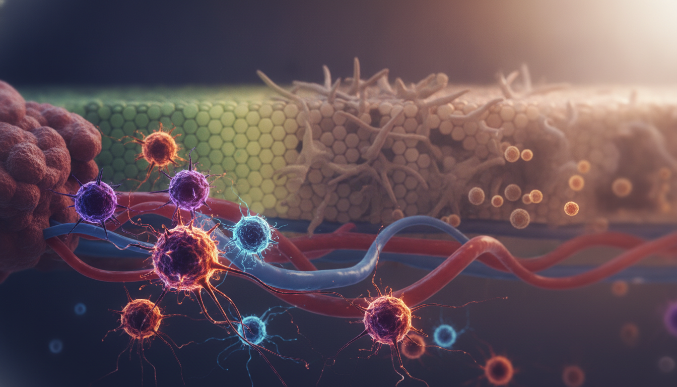
Metastatic Cancer Tracking
Markers of Epithelial-Mesenchymal Transition (EMT)
The regulation of the mesenchymal to epithelial transition is a sophisticated biological mechanism that allows stationary cells to become mobile. During this shift, cells lose their structural integrity and gain the capacity to infiltrate surrounding tissues. Researchers track this transformation by monitoring stem-cell-like markers such as SOX-2, OKT-4, and NANOG.
These markers provide insight into the plasticity of cancer cells. Additionally, MUC-1 is frequently analysed to assess the potential for further spread. By observing these changes, specialist healthcare providers can better predict the behaviour of aggressive tumours.
The Significance of EPCAM and PanCK
Identifying circulating tumour cells requires precise tools, and the epithelial marker profile is central to this effort. Proteins such as EPCAM and PanCK are essential for identifying cells that have entered the circulation. When a cell is identified as EPCAM+ve, it confirms the presence of epithelial-derived cancer cells in the blood.
Other critical proteins, such as C-MET and CD31, help in evaluating the vascularisation and invasive potential of these cells. Utilising these markers allows for a more comprehensive view of how cancer cells adapt to new environments. This detailed monitoring is a cornerstone of modern, personalised private oncology care in New Zealand.
At NatuneHeal, advanced oncology counts, tracing, onconomics and oncotrail, identification of circulating tumour cells and stem cells, as well as a wide variety of cancer markers are readily available to patients for a more detailed and focused approach to their health.
Balancing GSH and GSSG Levels
Managing oxidative stress is a cornerstone of integrative care, particularly when the body is under the strain of cancer therapies and treatments. The balance between GSH (reduced glutathione) and GSSG (oxidised glutathione) is a critical indicator of cellular health. When this ratio is disrupted, cells struggle to neutralise harmful free radicals.
Monitoring gamma-glutamyltransferase levels provides further clarity on how the body manages these antioxidant reserves. Maintaining this delicate balance is essential for improving cellular resilience and supporting the body’s natural recovery processes. Patients who focus on these markers often report feeling more supported throughout their therapy and treatment cycles.
The Role of Eicosanoid Related Proteins
The body’s inflammatory response is heavily influenced by the presence of specific signalling molecules. An eicosanoid related protein can act as a mediator in these pathways, affecting how tissues respond to injury or disease. Understanding these proteins helps clinicians see the broader picture of systemic inflammation.
Other markers, such as cytochrome C and P180, offer insights into mitochondrial health and protein synthesis. Furthermore, tracking a preprotein or identifying a specific proto-oncogene or fusion protein can reveal how cancer cells attempt to bypass normal regulatory mechanisms. When combined with the analysis of a repair related gene, these insights allow for a more comprehensive strategy to manage the tumour microenvironment effectively.
Navigating Cancer Prognosis and Therapy-Treatment Adjustments
Managing your health journey after a cancer diagnosis requires a clear roadmap for interpreting complex clinical data. By understanding the information provided by your laboratory reports, with the assistance of your specialists, you can feel more confident in the decisions made regarding your cancer prognosis.
Interpreting Test Results for Clinical Decisions
Only some specialist health providers rely on specific blood markers to gauge how your body is responding to therapy or treatment. These indicators provide a snapshot of your health, helping clinicians to determine whether a current health plan is working effectively or if a change is needed.
It is perfectly normal to feel overwhelmed when looking at technical laboratory reports. Always remember that these numbers are just one part of your broader health story, and they should be discussed in the context of your overall well-being.
When to Repeat Oncology Testing
Consistent cancer progression monitoring is vital for staying ahead of any changes in your condition. Repeating tests at scheduled intervals allows your specialists to identify trends rather than reacting to a single, isolated result.
If you notice new symptoms or if your clinical status shifts, do not hesitate to ask your Specialist Health Practitioner about the timing of your next assessment. Proactive testing ensures that your care plan remains as precise and responsive as possible.
Collaborating with Your Oncology Specialist Practitioner in New Zealand
Building a strong partnership with your healthcare providers in New Zealand is the foundation of successful therapy and treatment. Open communication allows you to discuss potential adjustments, such as the introduction of a proteasome inhibitor, with clarity and trust.
Your support team is there for you, so feel empowered to ask questions about your options. Working together ensures that your therapy and treatment remains tailored to your unique needs, helping you navigate your recovery with the best possible support system.
Conclusion
Modern medicine offers powerful tools to help you manage your wellbeing with confidence. Regular oncology testing to track cancer progression and tailor or customise therapy and treatment serves as a vital pillar for those seeking personalised care in New Zealand.
You gain significant clarity by utilising advanced biomarkers and genetic insights. These diagnostic methods allow you to monitor disease activity with remarkable precision. Working closely with your clinical team ensures that your therapy and treatment plan remains effective and responsive to your unique needs.
Proactive engagement with your health remains the best strategy for navigating complex health challenges. Consistent oncology testing to track cancer progression provides the data necessary for informed decision-making. You hold the power to shape your path forward by staying updated on these innovative diagnostic advancements.
We encourage you to discuss these options with your specialist healthcare providers to ensure your care aligns with your goals. Your commitment to understanding these tools makes a meaningful difference in your long-term health outcomes. Take charge of your journey today by prioritising the insights that matter most to your recovery.
FAQ
What is the primary benefit of oncology testing tracking cancer progression for patients in New Zealand?
The main advantage of oncology testing tracking cancer progression is the ability to monitor the disease with incredible precision. By using advanced pathology testing, clinicians can observe how a tumour responds to treatment in real-time. This proactive approach to tumour monitoring allows for timely adjustments to therapies, ensuring that the treatment remains effective as the neoplasmatic cells evolve.
How do Circulating Tumour Cells (CTCs) assist in cancer progression monitoring?
Circulating tumour cells count (CTCs) is a breakthrough in metastatic cancer tracking. These cells break away from the primary tumour and enter the bloodstream. Through malignant cell isolation and techniques like flow cytometry and immunophenotyping, we can analyse these cells to predict potential metastasis before it shows up on traditional scans. This is vital for early cancer diagnosis and long-term management.
What is the difference between Oncology counts and tracing tests?
Oncology counts are specifically designed to provide accurate CTC counts, helping to quantify the current systemic load of the disease. In contrast, Oncology tracing provides a deeper oncology biomarkers analysis, identifying the metastatic potential of the cells and distinguishing between standard CTCs and circulating stem cells count (CSCs). Oncology tracing also looks for specific markers such as CD34, CD133, and CD44 to assess the risk of spread.
Which blood markers are most commonly used for oncology screening?
Several blood markers are essential for different malignancies. For instance, CEA (carcinoembryonic antigen) is widely used in colorectal cancer screening, while PSA (prostate specific antigen) and acid prostatic phosphatase are standard for prostate cancer. Other specialised markers include thyroglobulin and calcitonin for thyroid cancer, and CA19.9, CA50, or CA15.3 for various adenocarcinomas and breast cancer.
How do Onconomics and Oncotrail provide personalised oncology insights?
Onconomics and Oncotrail utilise malignant cell culture to evaluate how a patient’s specific cancer cells respond to various natural therapies and conventional treatments. This includes testing chemosensitivity to biological modifier substances and conventional agents. These tests also examine the effectiveness of tyrosine kinase inhibition and proteasome inhibitors, allowing for a highly personalised health plan based on the tumour's unique biological profile.
What role do genetic mutations like VHL and BCR-ABL play in cancer prognosis?
Genetic mutations are key drivers of tumour growth monitoring. For example, the VHL mut is a primary indicator in renal carcinoma, while the BCR-ABL fusion protein is a hallmark of certain leukaemias. Monitoring these, along with tumour suppressor genes like PTEN and alk, helps clinicians identify resistant phenotype markers and predict the likely cancer prognosis, allowing for more aggressive or targeted interventions when necessary.
Can you explain the significance of CD markers in hematologic malignancies?
CD markers (Cluster of Differentiation) are proteins found on the surface of cells that help identify their origin. In hematologic malignancy markers analysis, we look for CD20, CD52, and CD33 to categorise lymphoma and myeloma. Markers like CD15 and CD30 help distinguish specific cell types, while CD34 and CD117 are often used to identify cells of myeloid cell origin or hematological stem cell and blast cell markers.
What markers are specific to lung cancer and neuroendocrine tumours?
For lung cancer, clinicians often track CYFRA 21.1 and specific neural enolase (SNE). These are particularly useful for identifying lung neuroendocrine malignancy. Additionally, markers like CA125 and mucin 1 (MUC-1) can provide further data on the epithelial nature of the tumour and its potential for migration invasion.
How does the tumour microenvironment influence angiogenesis and inflammation?
The microenvironment supports tumour growth through immunomodulation. Factors like VEGF, FGF, and PDGF drive angiogenesis (the growth of new blood vessels), while cytokines and growth factor receptors like EGF-R and IGF-R promote survival. Inflammatory pathways involving NFkb, COX2, and 5-LOX also play a significant role in maintaining the neoplasmatic cells and protecting them from oncogen apoptosis.
What is the Mesenchymal to Epithelial Transition (EMT) in metastatic cancer?
EMT is a process where cancer cells gain the ability to migrate and invade other tissues. We track this transition using markers such as EPCAM, PanCK, and vimentin. Monitoring the regulation of the mesenchymal to epithelial transition is crucial for understanding metastasis in aggressive cancers like metastatic melanoma, sarcoma, and colon cancer.
How does Advanced Oncology Testing and integrative approaches support patients during therapy and treatment?
Advanced Oncology Testing in New Zealand focuses on the patient’s internal biochemistry to support Natural therapy and/or conventional treatment. This includes monitoring GSH (glutathione) and GSSG levels to manage oxidative stress, as well as evaluating eicosanoid related proteins and glucose-6-phosphate-isomerase. By maintaining cellular health and immunomodulation, patients can often tolerate intensive treatments like tyrosine kinase inhibition more effectively while engaging in additional therapies of their choice.
What are immune checkpoint inhibitors like PD-1 and PD-L1?
PD-1, PD-L1, and PD-L2 are proteins that act as "brakes" on the immune system. Cancer cells often exploit these to hide from the body's natural defences. Testing for these markers helps determine if a patient is a good candidate for immunotherapy, which aims to release these brakes so the immune system can attack the malignant cell population directly.
When should a patient consider repeating their oncology testing?
Oncology testing should be repeated whenever there is a significant change in the clinical picture, such as the completion of a treatment cycle or if new symptoms arise. Regular cancer progression monitoring ensures that the oncology biomarkers analysis is up to date, allowing your specialist team to detect invasiveness or the development of a resistant phenotype early, ensuring your therapy and treatment strategy remains on the right track.